
Microscopy
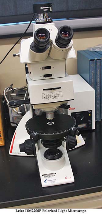
Leica DM2700P polarized light microscope.
A microscope consists mainly of an objective lens, ocular lens, lens tube, stage, and reflector. An object placed on the stage is magnified through the objective lens, and when the target is focused a magnified image can be observed through the ocular lens.
The laboratory uses several types of microscope, including low magnification stereozoom microscopes, higher powered microscopes that use polarized light to highlight certain optical characteristics of materials, and comparison microscopes, which pair two microscopes together to give a split view from each. Where higher magnification is necessary, the scanning electron microscope may be used to image the sample.
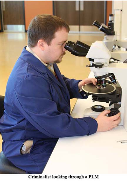
A criminalist at a polarized light microscope.
Fourier Transform Infrared Spectroscopy (FTIR)
FTIR is an instrumental technique used to identify compounds based on their infrared absorption characteristics. The spectra produced are used to conclusively identify compounds.
FTIR is used in the analysis of fibers, paint, tape, explosives, and substance identification.
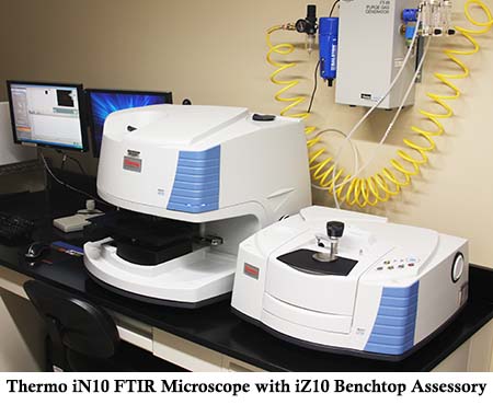
Thermo iN10 FTIR microscope with iZ10 benchtop accessory.
Scanning Electron Microscopy with Energy Dispersive Spectrometry (SEM/EDS)
The scanning electron microscope images extremely small particles using an electron beam that scans across the particle. Characteristic x-rays given off during scanning allow the energy dispersive spectrometer to determine the elemental makeup of the particle.
SEM/EDS is used in the analysis of gunshot residue, explosives, tape, soil, paint, and substance identification.
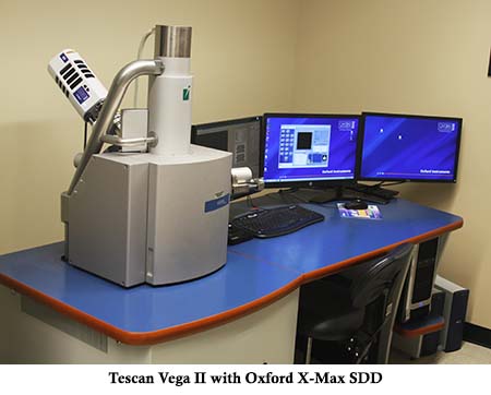
Tescan Vega II with Oxford X-Max SDD.
Gas Chromatography / Mass Spectrometry (GC/MS)
GC/MS combines two instrumental techniques. The gas chromatograph separates a mixture into its component parts and delivers them to the mass spectrometer, which breaks the molecules into ions and records the resulting spectrum. That spectrum conclusively identifies the compound.
GC/MS is used in the analysis of fire debris, explosives, paint, tape, and substance identification.
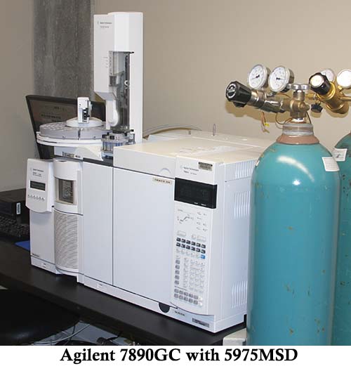
Agilent 7890 GC with 5975 MSD.
Microspectrophotometry (MSP)
The microspectrophotometer is a powerful analytical tool used for the study and color comparison of paint chips, fibers, and glass.
Spectra in the visible region provide an objective measurement of color, allowing comparisons between materials that may be indistinguishable to the naked eye.
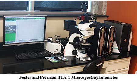
Foster and Freeman ffTA-1 microspectrophotometer.
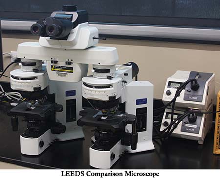
LEEDS comparison microscope.
X-ray Fluorescence Spectrometry (XRF)
XRF is used to determine the elemental composition of a sample. The analyzer measures the fluorescent x-rays emitted when the sample is excited by a high energy x-ray source. Those fluorescent x-rays are collected and plotted as a spectrum.
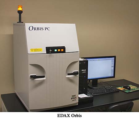
EDAX Orbis.
Raman Spectroscopy
Raman microscopy is a light scattering technique. When light from a high intensity laser interacts with a sample, most of the scattered light returns at the same wavelength as the laser source, known as Rayleigh scatter. A small percentage scatters at different wavelengths, depending on the chemical structure of the sample; this is Raman scattering.
The Raman microscope measures that scattering and plots it as a spectrum.
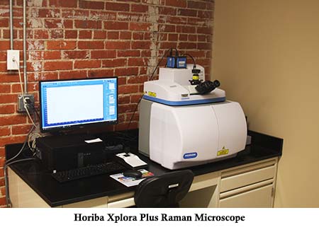
Horiba XploRA Plus Raman microscope.
Glass Refractive Index Measuring System (GRIM)
GRIM uses monochromatic light and temperature to graphically plot the refractive index of glass. Those refractive index values can be compared against a known glass sample to determine whether the glasses could have come from the same source.
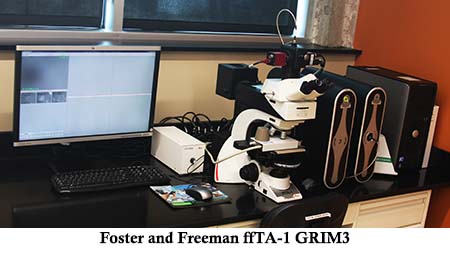
Foster and Freeman ffTA-1 GRIM3.